
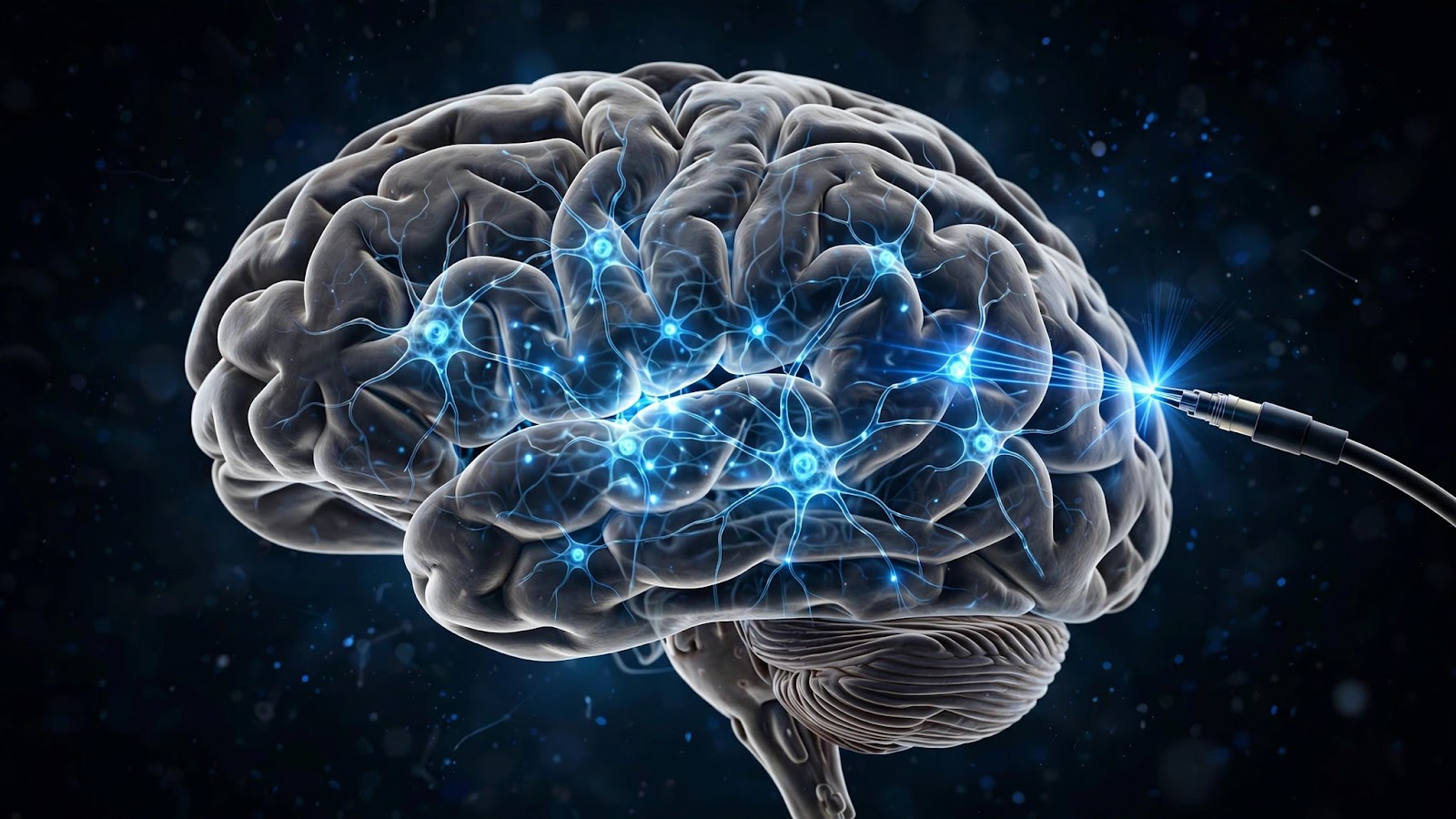
On the morning of October 5, 2026, the Nobel Assembly at the Karolinska Institutet in Stockholm announced the Nobel Prize in Physiology or Medicine. It was awarded jointly to Karl Deisseroth of Stanford University, Peter Hegemann of Humboldt University of Berlin, and Georg Nagel of the University of Würzburg, "for the discovery of light-gated ion channels and the development of optogenetics."
Few Nobel Prizes of recent decades have honored a tool as transformative as this one. Optogenetics lets scientists do something that sounds like science fiction: flip individual brain cells on and off with pulses of light, in living animals, with millisecond precision. Since its birth in 2005, the method has spread to thousands of laboratories, generated tens of thousands of papers, and rewritten what we know about memory, behavior, and disease. And it is now moving, cautiously but genuinely, toward the clinic.
The Algae That Saw the Light
The story begins not in a brain but in pond water. In the 1990s, the German biophysicist Peter Hegemann was studying Chlamydomonas reinhardtii, a single-celled green alga that swims toward light. The alga could sense light with remarkable speed, yet the protein doing the sensing was a mystery.
Working with Georg Nagel, Hegemann tracked the phenomenon down to a new family of proteins embedded in the alga's eyespot: the channelrhodopsins. In papers published in 2002 and 2003, the pair showed that these proteins were something never before seen in nature — ion channels that open in direct response to light. When blue light strikes a channelrhodopsin, the protein changes shape within microseconds and a pore opens, letting positively charged ions flood into the cell. The alga uses this electrical jolt to steer its flagella. In essence, the alga had evolved a light switch for its own cells.
At the time, almost no one imagined that this pond organism's trick would one day illuminate the human brain.
What Exactly Are Light-Gated Ion Channels?
To appreciate the Nobel, it helps to understand what a channelrhodopsin actually is. Every neuron communicates with tiny electrical pulses. When a neuron "fires," charged particles — ions — rush across its membrane through specialized protein channels, creating a spike of electricity called an action potential.
A channelrhodopsin is a single protein that combines two jobs: it is a light sensor and an ion channel in one molecule. Tucked inside the protein is a molecule called retinal — the same light-catching chemical found in your own eyes. When a photon of blue light (around 470 nanometers) hits the retinal, the protein snaps open a pore. Sodium and calcium ions pour in, the neuron's voltage rises, and the neuron fires. Turn the light off, and the channel closes within milliseconds.
Other light-sensitive proteins were soon added to the toolbox. Halorhodopsin, borrowed from salt-loving microbes, pumps chloride ions inward under yellow light and silences neurons. Archaerhodopsin pushes protons outward to do the same job. So neuroscientists gained not just an on-switch but a complete remote control: blue light to excite, yellow light to inhibit — all operating at the speed of thought itself.
Deisseroth's Leap: From Algae Protein to Brain Tool
The decisive leap came from Karl Deisseroth, a psychiatrist and neuroscientist at Stanford who wanted to understand the malfunctioning circuits behind depression and other brain disorders. The existing tools were blunt: drugs wash over the whole brain, and electrodes stimulate every cell type near their tip. Deisseroth wanted a scalpel.
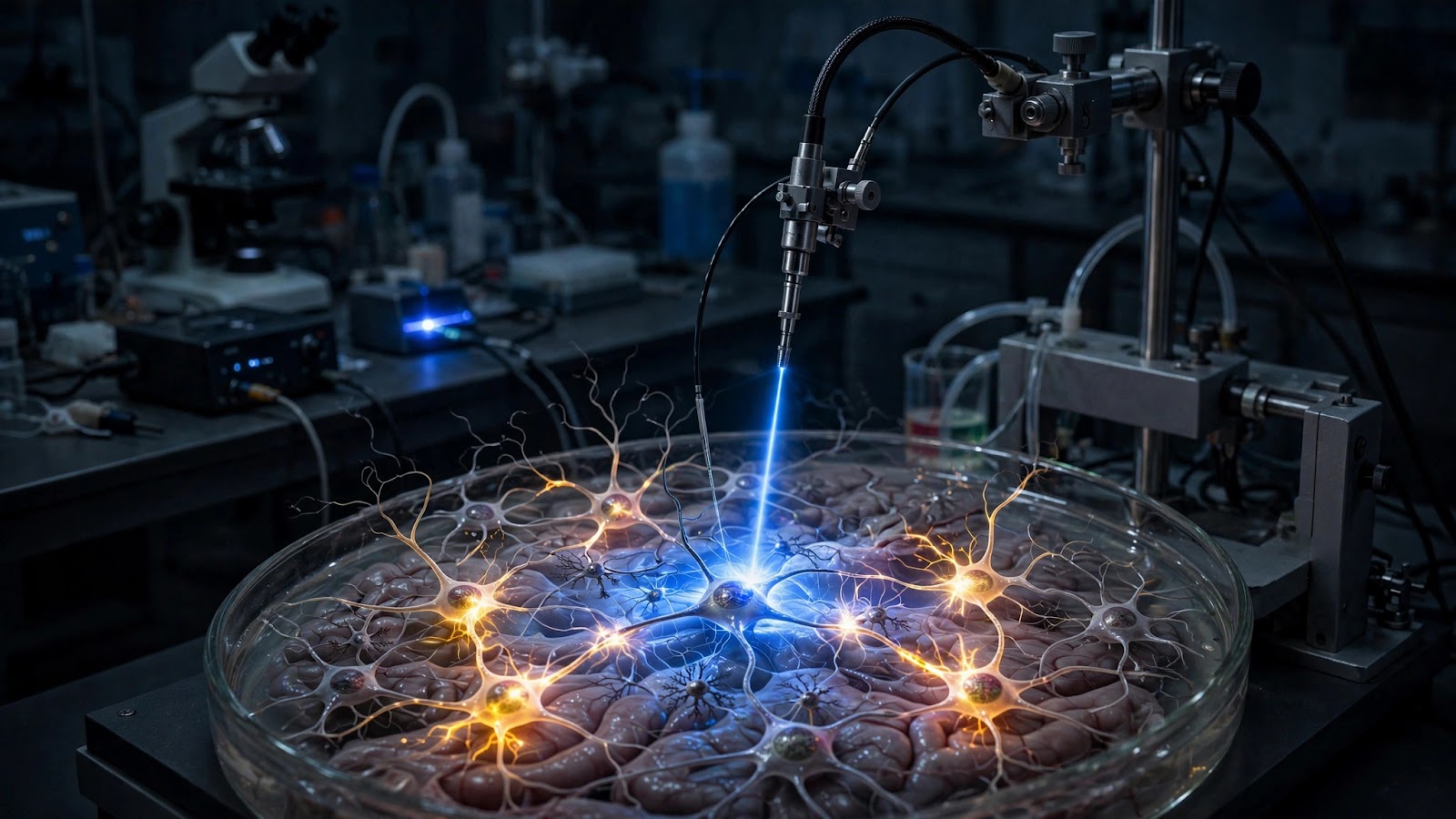
Reading the Hegemann–Nagel papers, he realized channelrhodopsin-2 could be that scalpel — if it worked in mammalian brain cells. With his student Ed Boyden and colleagues, he inserted the channelrhodopsin gene into cultured neurons, shone blue light on them, and watched the neurons fire in perfect lockstep with the light pulses. Their landmark paper, published in Nature Neuroscience in 2005, demonstrated "millisecond-timescale, genetically targeted optical control of neural activity."
That phrase — genetically targeted — was the real revolution. Because the light switch is a gene, scientists could aim it with genetic precision: only dopamine neurons, only the neurons carrying a particular receptor, only the cells active during a specific memory. Coupled with hair-thin fiber-optic cables implanted in the brain, researchers could now activate or silence exact cell types in awake, behaving animals. Deisseroth gave the method its name: optogenetics.
How Optogenetics Works, Step by Step
The modern optogenetics experiment follows a recipe that is conceptually simple, even if the engineering behind it is exquisite:
- Choose the cells. Researchers pick a genetic "address label" — a promoter that is active only in the neuron type they want to study, such as dopamine-producing neurons or the inhibitory interneurons of the cortex.
- Deliver the gene. A harmless, engineered virus carries the channelrhodopsin gene into the brain, where it installs itself only in cells carrying that address label. Those neurons now grow light-sensitive channels in their membranes.
- Install the light. A fiber-optic filament thinner than a human hair is implanted above the target region and connected to a laser or LED.
- Run the experiment. Flashes of blue light switch the chosen neurons on; yellow light switches them off. Cameras and sensors record what the animal does — and which circuits drive it.
Because the effect is reversible and instantaneous, a single animal can serve as its own control: light on, the behavior appears; light off, it vanishes — causation, not just correlation.
What Optogenetics Has Revealed About the Brain
Switching Memories On and Off
One of the most startling demonstrations came from memory research. In 2012, scientists at MIT tagged the exact hippocampus neurons that were active while a mouse formed a fear memory — the memory's physical trace, or engram — and installed channelrhodopsins in just those cells. Later, simply flashing blue light reactivated the memory: the mouse froze in fear even though nothing frightening was present. A follow-up study went further, activating two different engrams at once to implant what amounted to a false memory. The work showed, with unprecedented directness, that memories are stored in specific, reactivatable groups of cells — and that the line between a real memory and an implanted one is thinner than we thought.
Decoding Behavior: Hunger, Sleep, and Fear
Optogenetics has mapped the control panels of our most basic drives. Stimulating a tiny cluster of hunger neurons in the hypothalamus can make a fully fed mouse gorge itself within minutes; silencing the same cells makes a starving mouse stop eating. Specific circuits for thirst, aggression, mating behavior, and parental care have been pinpointed the same way.
Sleep has been another rich hunting ground. Researchers have identified the exact neuron populations that flip the brain between wakefulness and sleep, and shown how disrupting them fragments rest — findings with direct human relevance, since modern life is quietly eroding our nightly recovery (explored in our deep dive on how chronic sleep loss reshapes the brain).

Untangling the Circuits of Disease
For psychiatry and neurology, optogenetics has been nothing short of a circuit-diagram generator. In Parkinson's disease models, selectively driving or silencing pathways through the basal ganglia reproduces or relieves movement symptoms, clarifying exactly which connections go wrong. In depression research, activating reward-circuit neurons produces rapid antidepressant-like effects in animals, while silencing them induces anhedonia — the inability to feel pleasure that is a hallmark of human depression. These circuit maps are guiding new therapies, including precisely targeted deep-brain stimulation, and they complement practical strategies for maintaining mental health by revealing the biology those strategies must ultimately engage.
From Lab Bench to Bedside: Therapeutic Horizons
Restoring Vision
The most advanced clinical application is also the most poetic: giving light back to the blind. In diseases like retinitis pigmentosa, the eye's light-sensing photoreceptors die, but the downstream retinal neurons often survive. Optogenetic therapy aims to turn those surviving cells into replacement photoreceptors by delivering channelrhodopsin genes to them in a one-time injection.
In 2021, researchers reported in Nature Medicine that a blind patient with retinitis pigmentosa, treated with an optogenetic therapy and wearing light-amplifying goggles, had regained partial vision — able to perceive and count objects on a table. Several companies now have optogenetic vision therapies in clinical trials, and engineers are designing ever more sensitive opsins so that patients may one day need no goggles at all.
Treating Neurological and Psychiatric Disorders
Beyond vision, researchers are exploring optogenetic approaches to epilepsy — silencing seizure-prone circuits the instant abnormal activity begins, in a closed loop — as well as to Parkinson's disease, chronic pain, and even heart rhythm disorders, since heart muscle cells can be fitted with light switches too. These remain in animal studies for now, but the trajectory is clear: from observation tool to intervention.
The Hard Problems Still Ahead
Serious obstacles remain. Delivering genes safely to the human brain is harder than delivering them to a mouse's; the immune system may attack the foreign proteins; and blue light barely penetrates tissue, which is why scientists are engineering red-shifted opsins that respond to longer wavelengths that travel deeper. New light-delivery tricks — ultra-thin implants and even nanoparticles that convert infrared light into visible light inside the brain — are part of the race, an effort closely tied to how nanotechnology is reshaping medicine.
Why This Nobel Matters
Nobel Prizes sometimes reward discoveries and sometimes reward tools. Optogenetics is both. Hegemann and Nagel found a molecule that nature invented for an alga's eyespot; Deisseroth saw in it the answer to neuroscience's deepest methodological hunger — the ability to test, cell by cell, what each part of the brain actually does.
Before optogenetics, neuroscientists mostly watched the brain and correlated. After it, they could intervene — switching defined neurons on and off in a living, behaving animal and watching the result. That shift from correlation to causation is why the technique spread to thousands of laboratories and why its citation counts now rival those of PCR and CRISPR.
The deeper significance is philosophical as well as medical. Every time a flash of light conjures a memory, stills a seizure circuit, or returns a glimmer of sight to a blind eye, it confirms a radical idea: that the mind's most mysterious phenomena — memory, mood, perception — are the activity of identifiable cells, and are therefore, in principle, understandable and healable. That is the promise the Nobel Assembly recognized on October 5, 2026. The light switch is on, and neuroscience will never work in the dark again.